

 收藏
收藏
| 规格 | 价格 | 期货 | 数量 |
|---|
|
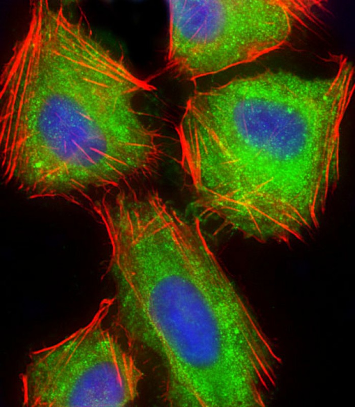
Immunofluorescent analysis of 4% paraformaldehyde-fixed, 0.1% Triton X-100 permeabilized U-2 OS (human osteosarcoma cell line) cells labeling ARPC5 with BD-PB5132 at 1/25 dilution, followed by Dylight® 488-conjugated goat anti-rabbit IgG secondary antibody at 1/200 dilution (green). Immunofluorescence image showing cytoplasm and weak nucleus staining on U-2 OS cell line. Cytoplasmic actin is detected with Dylight® 554 Phalloidin at 1/100 dilution (red).The nuclear counter stain is DAPI (blue). |
|
All lanes : Anti-ARPC5 Antibody (Center) at 1:2000 dilution Lane 1: THP-1 whole cell lysate Lane 2: HL-60 whole cell lysate Lane 3: Hela whole cell lysate Lane 4: MCF-7 whole cell lysate Lane 5: Human spleen lysate Lane 6: Mouse brain lysate Lane 7: Rat brain lysate Lysates/proteins at 20 µg per lane. Secondary Goat Anti-Rabbit IgG, (H+L), Peroxidase conjugated at 1/10000 dilution. Predicted band size : 16 kDa Blocking/Dilution buffer: 5% NFDM/TBST. |
 电话咨询
电话咨询
 在线咨询
在线咨询
 QQ
QQ
 二维码
二维码
 扫码二维码
扫码二维码