 |
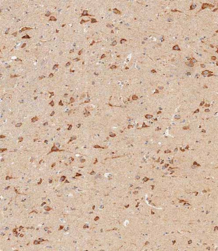
Immunohistochemical analysis of paraffin-embedded human brain tissue using BD-PB4645 performed on the Leica® BOND RXm. Tissue was fixed with formaldehyde at room temperature; antigen retrieval was by heat mediation with a EDTA buffer (pH9. 0). Samples were incubated with primary antibody(1:1000) for 1 hours at room temperature. A undiluted biotinylated CRF Anti-Polyvalent HRP Polymer antibody was used as the secondary antibody. |
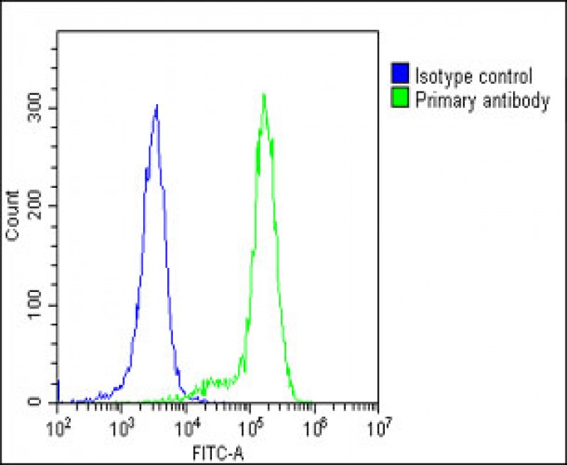 |
Overlay histogram showing K562 cells stained with BD-PB4645(green line). The cells were fixed with 2% paraformaldehyde (10 min) and then permeabilized with 90% methanol for 10 min. The cells were then icubated in 2% bovine serum albumin to block non-specific protein-protein interactions followed by the antibody for 60 min at 37ºC. The secondary antibody used was Goat-Anti-Rabbit IgG, DyLight® 488 Conjugated Highly Cross-Adsorbed at 1/200 dilution for 40 min at 37ºC. Isotype control antibody (blue line) was rabbit IgG1 (1μg/1x10^6 cells) used under the same conditions. Acquisition of >10, 000 events was performed. |
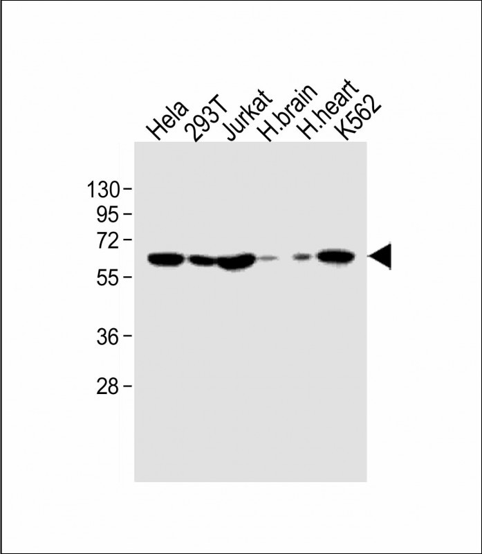 |
All lanes : Anti-IFNAR1 Antibody (Center) at 1:1000 dilution
Lane 1: Hela whole cell lysate
Lane 2: 293T whole cell lysate
Lane 3: Jurkat whole cell lysate
Lane 4: human brain lysate
Lane 5: human heart lysate
Lane 6: K562 whole cell lysate
Lysates/proteins at 20 µg per lane.
Secondary
Goat Anti-Rabbit IgG, (H+L), Peroxidase conjugated at 1/10000 dilution.
Predicted band size : 64 kDa
Blocking/Dilution buffer: 5% NFDM/TBST. |


 收藏
收藏

 电话咨询
电话咨询
 在线咨询
在线咨询
 QQ
QQ
 二维码
二维码
 扫码二维码
扫码二维码