 |
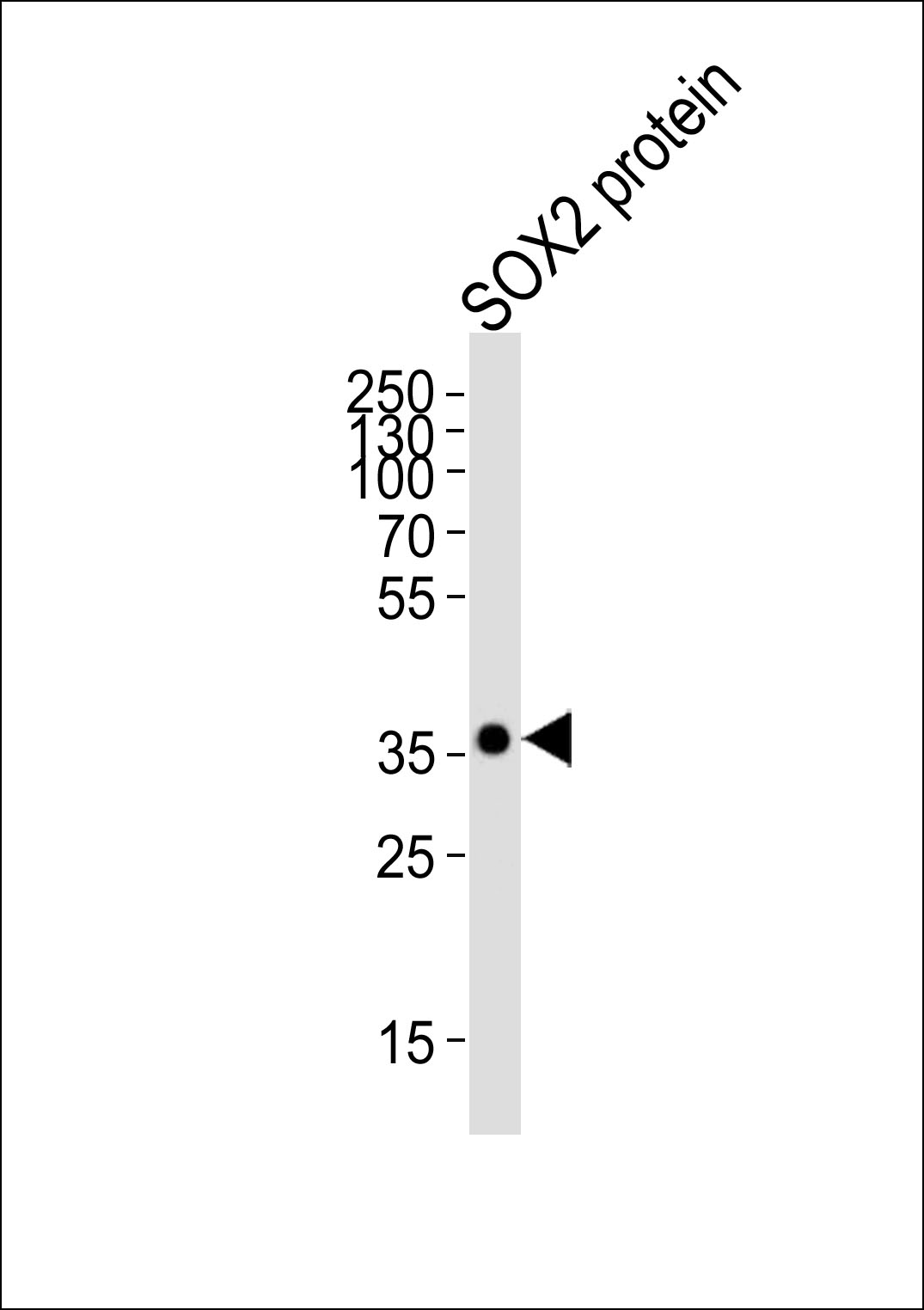
Western blot analysis of lysate from SOX2 protein, using SOX2 Antibodyat 1:3000 dilution was used as the secondary antibody. Lysate at 20μg. |
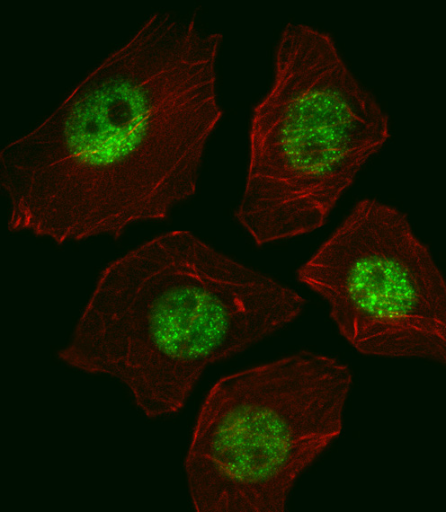 |
Fluorescent image of A549 cell stained with SOX2 Antibodywas used (1:400, 50 min at 37℃.Cytoplasmic actin was counterstained with Alexa Fluor® 555 (red) conjugated Phalloidin (7units/ml, 1 h at 37℃.SOX2 immunoreactivity is localized to Nucleus significantly. |
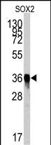 |
Western blot analysis of SOX2 Antibody by SOX2 recombinant protein. SOX2(arrow) was detected using the purified Mab. |
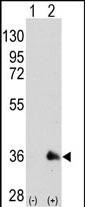 |
Western blot analysis of SOX2 (arrow) using mouse monoclonal SOX2 antibody either nontransfected (Lane 1) or transiently transfected with the SOX2 gene (Lane 2) |
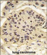 |
Formalin-fixed and paraffin-embedded human lung carcinoma tissue reacted with SOX2 Antibody , which was peroxidase-conjugated to the secondary antibody, followed by DAB staining. This data demonstrates the use of this antibody for immunohistochemistry; clinical relevance has not been evaluated. |
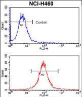 |
Flow cytometric analysis of NCI-H460 cells using SOX2 Monoclonal Antibody (bottom histogram) compared to a negative control cell (top histogram). PE-conjugated goat-anti-mouse secondary antibodies were used for the analysis. |
 |
Immunohistochemical analysis of paraffin-embedded Human Lung squamous cell carcinoma section using Pink1. BD-PB3860 was diluted at 1:100 dilution. A undiluted biotinylated goat polyvalent antibody was used as the secondary, followed by DAB staining. |


 收藏
收藏

 电话咨询
电话咨询
 在线咨询
在线咨询
 QQ
QQ
 二维码
二维码
 扫码二维码
扫码二维码