SNAI 1 Rabbit Polyclonal Antibody

货号: BD-PT4351
收藏
规格 价格 期货 数量
  • 50μl
    1100.00
    现货
    100μl
    2000.00
    现货

产品基本信息

  • 产品货号 BD-PT4351
  • 别名 SNAI1; SNAH; Zinc finger protein SNAI1; Protein snail homolog 1; Protein sna
  • 产品名称 SNAI 1 Rabbit Polyclonal Antibody
  • 类别 抗原抗体
  • 基因名称 SNAI1
  • 蛋白名称 Zinc finger protein SNAI1(snail)
  • Clonality Polyclonal
  • 推荐应用 WB,IP,IHC-P,IF-P,IF-F,IF-ICC,ELISA
  • 反应种属 Human,Mouse,Monkey
  • 浓度 1 mg/ml
  • 存储缓冲液 Liquid in PBS containing 50% glycerol, 0.5% BSA and 0.02% New type preservative N.
  • Human Gene ID 6615
  • Human Gene Link http://www.ncbi.nlm.nih.gov/sites/entrez?db=gene&term=6615
  • Human Swissprot Link http://www.uniprot.org/uniprotkb/O95863/entry
  • Mouse Gene ID 20613
  • Mouse Gene Link http://www.ncbi.nlm.nih.gov/sites/entrez?db=gene&term=20613
  • Mouse Swissprot No. Q02085
  • Mouse Swissprot Link http://www.uniprot.org/uniprot/Q02085
  • 免疫原 The antiserum was produced against synthesized peptide derived from human SNAI1. AA range:215-264
  • 特异性 SNAI 1 Polyclonal Antibody detects endogenous levels of SNAI 1 protein.
  • 稀释度 WB 1:500-1:2000, IHC-P 1:100-1:300, IF-P/IF-F/IF-ICC 1:200-1:1000, ELISA 1:5000.Not yet tested in other applications.
  • 预测分子量 29kDa
  • 运输及保存条件 -20°C/1 year
  • 宿主 Rabbit
  • 同种型 Rabbit,IgG
  • 背景介绍 snail family transcriptional repressor 1(SNAI1) Homo sapiens The Drosophila embryonic protein snail is a zinc finger transcriptional repressor which downregulates the expression of ectodermal genes within the mesoderm. The nuclear protein encoded by this gene is structurally similar to the Drosophila snail protein, and is also thought to be critical for mesoderm formation in the developing embryo. At least two variants of a similar processed pseudogene have been found on chromosome 2. [provided by RefSeq, Jul 2008],
  • 组织表达 Expressed in a variety of tissues with the highest expression in kidney. Expressed in mesenchymal and epithelial cell lines.
  • 细胞定位 Nucleus . Cytoplasm . Once phosphorylated (probably on Ser-107, Ser-111, Ser-115 and Ser-119) it is exported from the nucleus to the cytoplasm where subsequent phosphorylation of the destruction motif and ubiquitination involving BTRC occurs. .
  • 信号通路 Adherens_Junction;
  • 功能 function:Seems to be involved in embryonic mesoderm formation. Binds to 3 E-boxes of the E-cadherin gene promoter and represses its transcription.,similarity:Belongs to the snail C2H2-type zinc-finger protein family.,similarity:Contains 4 C2H2-type zinc fingers.,tissue specificity:Expressed in a variety of tissues with the highest expression in kidney.,
  • 纯化 The antibody was affinity-purified from rabbit antiserum by affinity-chromatography using epitope-specific immunogen.

图片

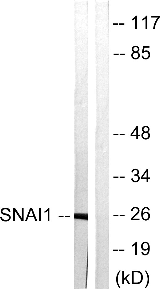
Western blot analysis of lysates from HT29 cells, using SNAI1 Antibody. The lane on the right is blocked with the synthesized peptide.
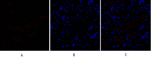
Immunofluorescence analysis of rat-heart tissue. 1,SNAI 1 Polyclonal Antibody(red) was diluted at 1:200(4°C,overnight). 2, Cy3 labled Secondary antibody was diluted at 1:300(room temperature, 50min).3, Picture B: DAPI(blue) 10min. Picture A:Target. Picture B: DAPI. Picture C: merge of A+B
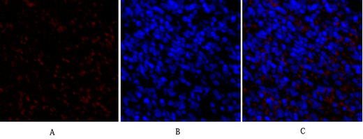
Immunofluorescence analysis of rat-kidney tissue. 1,SNAI 1 Polyclonal Antibody(red) was diluted at 1:200(4°C,overnight). 2, Cy3 labled Secondary antibody was diluted at 1:300(room temperature, 50min).3, Picture B: DAPI(blue) 10min. Picture A:Target. Picture B: DAPI. Picture C: merge of A+B
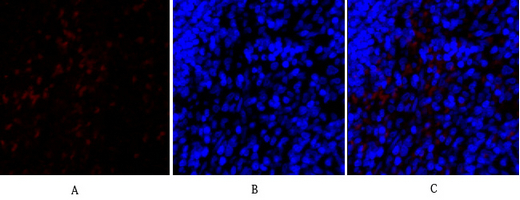
Immunofluorescence analysis of rat-spleen tissue. 1,SNAI 1 Polyclonal Antibody(red) was diluted at 1:200(4°C,overnight). 2, Cy3 labled Secondary antibody was diluted at 1:300(room temperature, 50min).3, Picture B: DAPI(blue) 10min. Picture A:Target. Picture B: DAPI. Picture C: merge of A+B
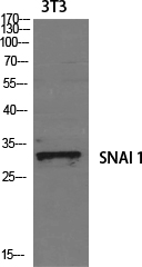
Western Blot analysis of various cells using SNAI 1 Polyclonal Antibody diluted at 1:1000 cells nucleus extracted by Minute TM Cytoplasmic and Nuclear Fractionation kit (SC-003,Inventbiotech,MN,USA).
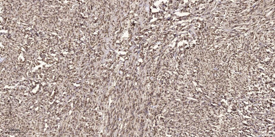
Immunohistochemical analysis of paraffin-embedded human small intestinal carcinoma tissue. 1,primary Antibody was diluted at 1:200(4° overnight). 2, Sodium citrate pH 6.0 was used for antigen retrieval(>98°C,20min). 3,Secondary antibody was diluted at 1:200

产品详情

文献引用 (0)

产品问答

内容已折叠

相关产品

内容已折叠

最新浏览的产品

如果您对产品或服务感兴趣,可通过以下方式联系我们

  • 在下方表单 填写需求描述给我们
  • 点击页面右侧“在线咨询” 工具快速咨询
  • 拨打免费电话 400-921-8278
  • 发送邮件至邮箱 sales@biodragon.cn
« »