|
Western blot analysis of lysates from HCT-116 cells, using KLF4 Rabbit mAb. The HRP-conjugated Goat anti-Rabbit IgG antibody was used to detect the antibody. |
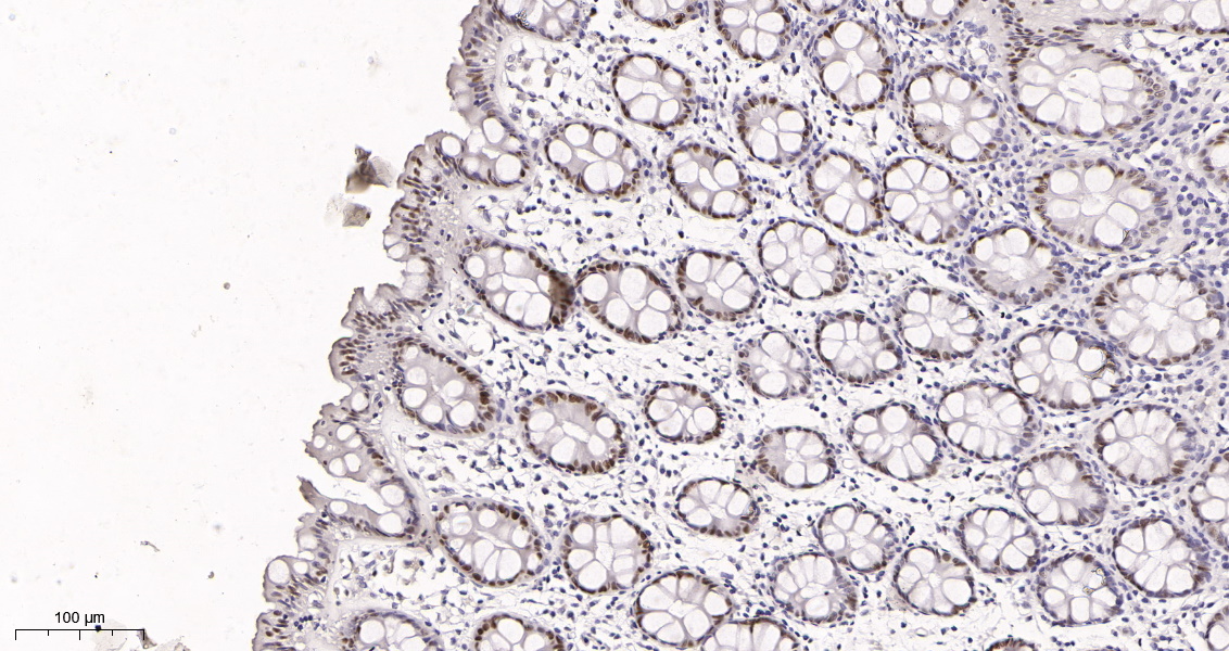 |
Immunohistochemical analysis of paraffin-embedded human colon tissue. 1, primary Antibody was diluted at 1:200(4℃,overnight). 2, EDTA pH 9.0 was used for antibody retrieval(>98℃,20min). 3,Secondary antibody was diluted at 1:200(room tempeRature, 30min). |
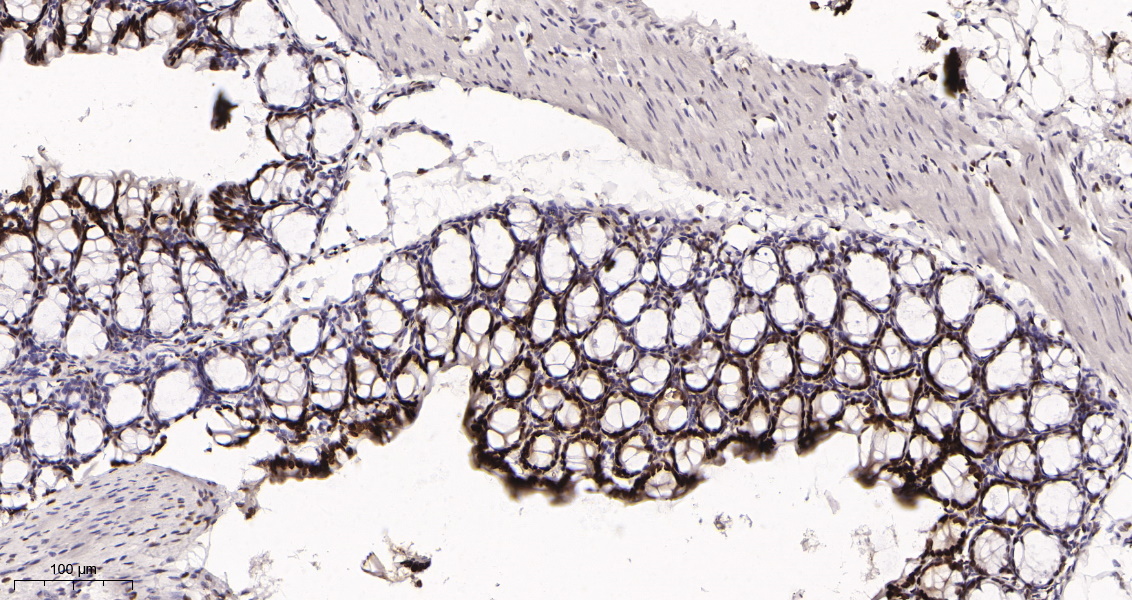 |
Immunohistochemical analysis of paraffin-embedded mouse colon tissue. 1, primary Antibody was diluted at 1:200(4℃,overnight). 2, EDTA pH 9.0 was used for antibody retrieval(>98℃,20min). 3,Secondary antibody was diluted at 1:200(room tempeRature, 30min). |
 |
Immunohistochemical analysis of paraffin-embedded rat colon tissue. 1, primary Antibody was diluted at 1:200(4℃,overnight). 2, EDTA pH 9.0 was used for antibody retrieval(>98℃,20min). 3,Secondary antibody was diluted at 1:200(room tempeRature, 30min). |


 收藏
收藏
 电话咨询
电话咨询
 在线咨询
在线咨询
 QQ
QQ
 二维码
二维码
 扫码二维码
扫码二维码